
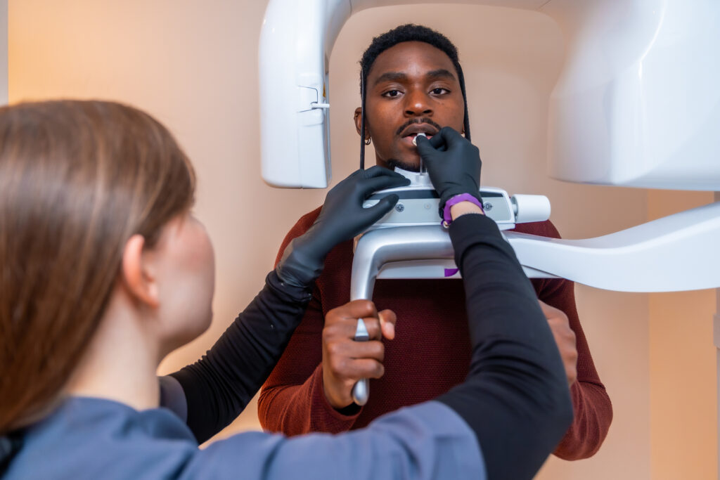
A CBCT 3D scan gives your dental team a complete, three-dimensional view of your jawbone, nerves, and surrounding anatomy before any implant work begins. That level of detail changes everything about how treatment is planned, because your dentist can measure bone density, map nerve pathways, and identify potential complications long before you sit in the chair for a procedure. Without this kind of imaging, implant planning relies on two-dimensional X-rays that flatten complex structures into a single plane, leaving critical questions unanswered until surgery day.
At Sopris Smiles, CBCT imaging is a standard part of how we approach dental implant restorations. Dr. Golnaz uses these scans to work collaboratively with oral surgery colleagues, ensuring each implant is planned for the most ideal positioning before placement ever begins. The result is a more precise, more predictable process from start to finish.
What a CBCT Scan Reveals That Traditional X-Rays Cannot
A traditional dental X-ray captures a flat, two-dimensional image. It can show the general shape of your jawbone and the location of your teeth, but it compresses everything into a single layer. Overlapping structures, hidden bone loss, and the exact position of the inferior alveolar nerve can all be obscured.
A CBCT scan, by contrast, produces a full three-dimensional reconstruction of your jaw. Your dental team can rotate the image, zoom into specific cross-sections, and measure bone width, height, and density at the exact site where an implant would be placed. A 2024 systematic review published by the National Institutes of Health confirmed CBCT provides accurate linear measurements for implant planning, making it an appropriate diagnostic tool for three-dimensional preoperative assessment.
Why Bone Dimensions Matter
Implants need a certain amount of healthy bone to anchor securely. If the bone is too narrow, too shallow, or too soft, the implant may not integrate properly. CBCT imaging lets your care team identify these limitations in advance so the treatment plan can be adjusted, whether through selecting a different implant size, choosing an alternative site, or coordinating bone grafting with a surgeon when necessary.
How CBCT Imaging Shapes the Implant Planning Process
Once a CBCT scan is captured, the data is loaded into planning software where your dentist can virtually position the implant within the three-dimensional model of your jaw. This step allows for precise angulation, depth selection, and spacing relative to adjacent teeth and nerves.
At our Englewood office, we pair CBCT scans with CEREC digital scanning technology to create a comprehensive picture of both your bone structure and your existing dental anatomy. When a case calls for it, this data can be used to create a personalized surgical guide for the oral surgeon, helping ensure the implant is positioned in the most ideal location for a long-lasting restoration.
Benefits of Digital Implant Planning
Here are some of the key advantages CBCT-guided implant planning offers over conventional methods:
- Precise bone measurements reduce the risk of nerve injury or sinus perforation during implant placement
- Three-dimensional visualization allows for more accurate implant sizing and angulation
- Collaborative planning between your restorative dentist and oral surgeon leads to better coordination
- Patients receive a clearer picture of their treatment timeline and what to expect at each stage
Each of these benefits contributes to a smoother process and a stronger foundation for the final crown, bridge, or other restoration.
The Collaborative Approach to Implant Restorations
Our office coordinates closely with oral surgeons who handle the surgical placement of implants. Dr. Golnaz’s role centers on the planning and restoration side, taking the CBCT scan, analyzing the data, and working with the surgeon to map out the precise location for each implant. After placement and healing, our team completes the process with a custom abutment and crown designed using CEREC same-day crown technology.
This team-based approach means no detail gets lost in translation. The surgeon knows exactly where the implant needs to go for the best possible cosmetic and functional result, and we can design the final restoration with confidence because the foundation was placed with precision.
Learn More About CBCT-Guided Implant Planning at Sopris Smiles
Every dental implant restoration at our Englewood office begins with a detailed evaluation, not guesswork. Dr. Golnaz uses CBCT 3D imaging and CEREC digital scanning to get a clear picture of your teeth, jaw, and bone before treatment begins. She also works closely with trusted oral surgery specialists to carefully plan each case. Along the way, she looks at more than just the missing tooth, considering how your implant will fit your bite, support your long-term oral health, and help you reach your goals for your smile.
If you’re thinking about dental implants and want to know how advanced imaging can improve your outcome, we’d love to walk you through the process. Contact our team today to schedule a consultation and see what CBCT-guided planning looks like for your smile.